
Stay up-to-date
Upcoming events
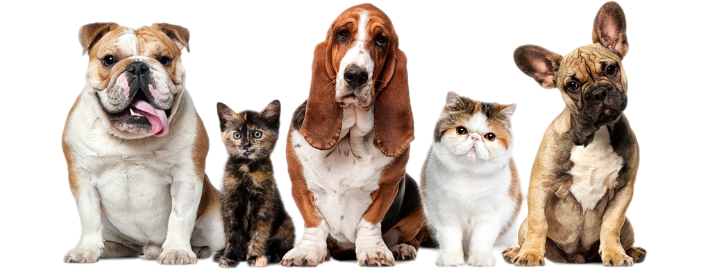
Veterinary Education & Training to further your career
We are offering Practical Education Workshops for Veterinarians
Expert educators
We’re passionate about ensuring our students succeed. Our instructors are diplomates and specialists in their fields so they can pass on their expertise and knowledge to our students
Online Courses
Gain hands-on experience in a virtual medical environment. Workshops provide you with the knowledge and skills at the comfort of your office, clinic, or home.
Global Classroom
A complete built-in social network for connecting with students. IEVS platform helps you connect with fellow veterinarians across the globe.
Latest news from IEVS
IEVS is a project of the ESAVS Office for Asia
European School for Advanced Veterinary Studies (ESAVS)
with more than 25 years experience in residential courses
Our Webinars
\Learnworlds\Codeneurons\Pages\ZoneRenderers\CourseCards
High quality veterinary continuous education for vets
Completing online continuing education courses is a great way to stay current on the latest techniques and procedures, as well as keeping current on the most important legislation and regulations.
Get the newsletter!
Stay updated | Stay current | Stay connected
Thank you!
Copyright © 2023